


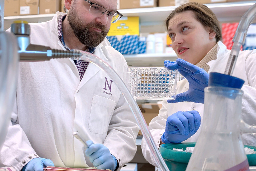
 Jason Miska, PhD, and Leah Billingham, PhD, read out enzymatic activity of microglia in cell cultures.
Photo: Ben Schamisso
Northwestern Medicine scientists have discovered that specialized immune cells within the glioblastoma tumor microenvironment metabolize fructose to suppress immune responses and promote tumor growth, according to a recent study published in the Proceedings of the National Academy of Sciences.
The findings suggest that blocking fructose metabolism in these cells may improve immunotherapy response and patient outcomes, according to Jason Miska, PhD, assistant professor of Neurological Surgery and senior author of the study.
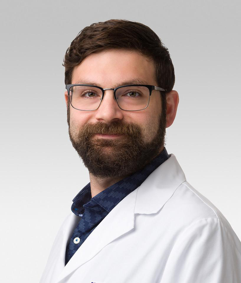 Jason Miska, PhD, assistant professor of Neurological Surgery, was senior author of the study published in the Proceedings of the National Academy of Sciences.
Glioblastoma is the most common and aggressive primary brain tumor in adults and has maintained a five-year survival rate of less than 7 percent, according to the National Brain Tumor Society. Glioblastoma is one of the most treatment-resistant brain tumors due to its tumor microenvironment, which includes immunosuppressive myeloid cells, immune cells that originate from bone marrow, and brain-resident microglia — immune cells of the brain and central nervous system.
Microglia have been shown to be crucial for the early stages of glioma growth and maintain unique metabolic and immunologic processes in glioblastoma compared to infiltrating myeloid cells. Microglia also express a unique fructose transporter, GLUT5, enabling them to transport and metabolize fructose. The role of microglial fructose metabolism in glioblastoma tumor progression, however, has remained poorly understood, Miska said.
“There is more fructose in cerebral spinal fluid than there is in the plasma. The reasons for this are unknown, but what is known is that microglia express a lot of the transporter to metabolize it. So, we were curious about what this metabolism meant for brain tumors,” said Miska, who is also a member of the Robert H. Lurie Comprehensive Cancer Center of Northwestern University.
In mouse models of glioblastoma, the scientists used a combination of flow cytometry, bulk RNA- sequencing, and single-cell RNA- sequencing techniques to profile microglia, macrophages and glioma cell lines from the tumors and surrounding tissue.
This analysis not only confirmed that microglia uniquely express GLUT5 but also showed that microglia are the only immune cells in the glioblastoma microenvironment capable of metabolizing fructose.
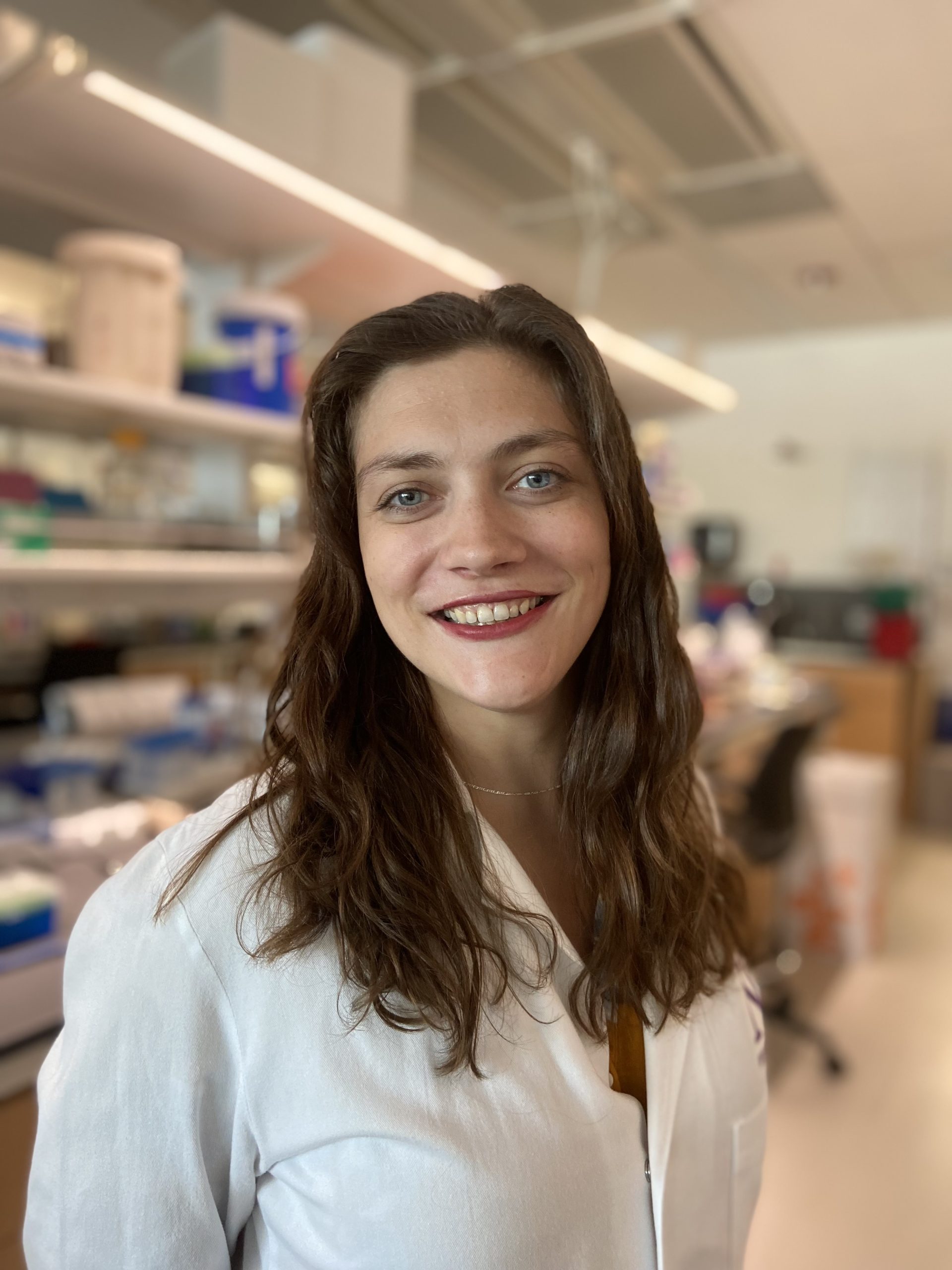 Leah Billingham, PhD, a postdoctoral fellow in Miska laboratory, was co-first author of the study.
The scientists also found that deleting GLUT5 prevented tumor establishment in several glioma models, driven by a loss of fructose metabolism in microglia within the tumor microenvironment.
Furthermore, tumors in GLUT5 knockout mice demonstrated increased activation of innate and adaptive immune responses, including enhanced antigen presentation, clonal expansion (rapid division) of CD8+ T-cells, and increased cytokine production.
“This not only makes the microglia themselves more inflammatory, but it also causes those T-cells and B-cells that are in the tumor to be more activated and create more inflammatory molecules that we have shown are required for that rejection of brain tumors,” said Leah Billingham, PhD, a postdoctoral fellow in Miska laboratory and co-first author of the study. “This isn’t just solely the microglia doing something; this is an intricate interaction between the different parts of the immune system and how they are then impacting tumor rejection,” Billingham said.
The findings suggest microglial fructose metabolism is a key regulator of immune suppression in glioblastoma and may be a promising therapeutic target to improve immunotherapy response in patients.
Miska also noted the unique role of fructose in the brain compared to other organ systems: increased fructose consumption is associated with many inflammatory diseases, including colon cancer and diabetic neuropathy, but in the brain, fructose seems to instead suppresses inflammation.
“Fructose consumption is associated with so many bad inflammatory outcomes in patients. What’s interesting here is that in the brain, it seems to be working differently,” Miska said. “It still helps the brain tumor grow, but now we’re seeing something very different in the brain than we see in the rest of the body,” Miska said.
Going forward, Miska said his team aims to identify promising fructose transport inhibitors that can then be tested in preclinical trials.
“Once we can get our hands on something that is promising as a fructose transport inhibitor, we will then take it into preclinical stages where we add standard-of-care therapies for brain tumors or immunotherapies and see if we can sensitize them,” Miska said.
Co-authors include Tzu-yi Chia, Ian Olson, Ruochen Du, Hanxiao Wan and Joseph Duffy, students in the Driskill Graduate Program in Life Sciences (DGP); Shashwat Tripathi, a student in the Medical Scientist Training Program (MSTP); Nishanth Sadagopan, a third-year medical student; Alina Murphy, PhD, a postdoctoral fellow in the Lee-Chang laboratory; Irina Balyasnikova, PhD, professor of Neurological Surgery; Peng Zhang, PhD, assistant professor of Neurological Surgery; Atique Ahmed, PhD, the Allen Buckner Kanavel Professor of Neurosurgery; Catalina Lee-Chang, PhD, assistant professor of Neurological Surgery; Amy Heimberger, MD, PhD, the Jean Malnati Miller Professor of Brain Tumor Research and vice chair for research in the Department of Neurological Surgery; and Navdeep Chandel, PhD, the David W. Cugell, MD, Professor of Medicine in the Division of Pulmonary and Critical Care.
This work was supported by National Cancer Institute grants 5R01CA279686-03, P50CA221747, T32CA070085, R37CA258426, P30CA060553 and R37CA266487; Cancer Research Institute grants CR68036 and CR13733; National Institute of Neurological Disorders and Stroke grants R01NS122395 and NS120547; National Institute of General Medical Sciences grant 1DP2GM146337; MSKCC Cancer Center Support Grant P30CA008748; and National Institute of Allergy and Infectious Diseases grant 5T32AI134632.